

Muscle Types
Muscle tissue has four main properties: Excitability (ability to respond to stimuli), Contractibility (capacity to contract), Extensibility (ability to be stretched without tearing) and Elasticity (ability to return to its standard shape). Based on specific structural and functional characteristics, muscle tissue is classified into three types: cardiac, smooth and skeletal.
Cardiac
Cardiac muscle tissue forms the bulk of the wall of the heart. Like skeletal muscle tissue, it is striated (the muscle fibres contain alternating light and dark bands (striations) perpendicular to the long axes of the fibres). Unlike skeletal muscle tissue, contraction is usually not under conscious control (involuntary).
Smooth
Smooth muscle fibres are usually involuntary (not under conscious control) and nonstriated (smooth). Smooth muscle tissue is located in the walls of hollow internal structures such as blood vessels, the stomach, the intestines, and the urinary bladder. Smooth muscle tissue, like skeletal and cardiac muscle tissue, can undergo hypertrophy. Also, individual smooth muscle fibres, such as those in the uterus, retain their capacity for division and can grow by hyperplasia.
Skeletal
Skeletal muscle tissue is named for its location - attached to bones. It is striated; the fibres (cells) contain alternating light and dark bands (striations) perpendicular to the long axes of the fibres. Skeletal muscle tissue can be contracted or relaxed by conscious control (voluntary).
All skeletal muscle fibres are not alike in structure or function. For example, skeletal muscle fibres vary in colour depending on myoglobin content (myoglobin stores oxygen until needed by the mitochondria). Skeletal muscle fibres contract with different velocities, depending on their ability to split Adenosine Triphosphate (ATP). Faster contracting fibres have a greater ability to split ATP. Also, skeletal muscle fibres vary concerning the metabolic processes they use to generate ATP. They also differ in terms of the onset of fatigue. Based on various structural and functional characteristics, skeletal muscle fibres are classified into: Type I fibres, Type II B fibres and Type II A fibres.
FasciaThe following is the definition of Fascia provided by the Fascia Research Congress (2009)[2] "Fascia is the soft tissue component of the connective tissue system. It interpenetrates and surrounds muscles, bones, organs, nerves, blood vessels and other structures. Fascia is an uninterrupted, three-dimensional web of tissue that extends from head to toe, from front to back, and from interior to exterior. |
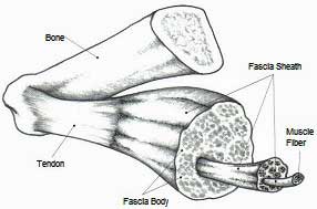 |
It is responsible for maintaining structural integrity, providing support and protection, and acting as a shock absorber. Fascia is essential in hemodynamic and biochemical processes and provides the matrix that allows for intercellular communication.
After an injury, the fascia creates an environment for tissue repair. Fascia can refer to dense planar fascial sheets (such as the Fascia lata) and joint capsules, organ capsules, muscular septa, ligaments, retinacula, aponeuroses, tendons, myofascia, neurofascia, and other fibrous collagenous tissues".
Langevin and Huijing (2009)[1] state that we need to be aware of possible ambiguities and misunderstandings stemming from different meanings of the word "Fascia" because the general meaning of the term can be so vague as to imply little more than some form of connective tissue. "Fascia" encompasses both loose and dense, superficial and deep, and multiple and single-layered connective tissues.
Myoglobin & Mitochondria
Myoglobin is a Protein with oxygen bound to it, thus providing an extra reserve of oxygen so that the muscle can maintain a high activity level for a longer time.
Mitochondria are known as the powerhouses of the cell. They act like a digestive system that takes in nutrients, breaks them down, and creates energy for the cell.
Type I Fibres
Type I fibres are red, split ATP at a slow rate, have a slow contraction velocity, are very resistant to fatigue and have a high capacity to generate ATP by oxidative metabolic processes. These fibres, also called slow-twitch or slow oxidative fibres, contain large amounts of myoglobin, mitochondria, and blood capillaries. Such fibres are found in large numbers in the neck's postural muscles.
Type II A Fibres
Type II A fibres are red, have a very high capacity for generating ATP by oxidative metabolic processes, split ATP rapidly, have a fast contraction velocity and are resistant to fatigue. These fibres also called fast twitch or fast oxidative fibres, contain vast amounts of myoglobin, many mitochondria and blood capillaries. Such fibres are infrequently found in humans.
Type II B Fibres
These fibres also called fast twitch or fast glycolytic fibres, contain a low content of myoglobin, relatively few mitochondria, relatively few blood capillaries and large amounts of glycogen. Type II B fibres are white, geared to generate ATP by anaerobic metabolic processes, unable to supply skeletal muscle fibres continuously with sufficient ATP, fatigues quickly, split ATP at a fast rate and has a rapid contraction velocity. Such fibres are found in large numbers in the muscles of the arms.
Characteristics of Muscle Types
The following table is adapted from Honeybourne et al. (1996)[3]
| Fibre Type | Type I fibres | Type II A fibres | Type II B fibres |
| Contraction time | Slow | Fast | Very Fast |
| Size of motor neuron | Small | Large | Very Large |
| Resistance to fatigue | High | Intermediate | Low |
| Activity Used for | Aerobic | Long-term anaerobic | Short term anaerobic |
| Force production | Low | High | Very High |
| Mitochondrial density | High | High | Low |
| Capillary density | High | Intermediate | Low |
| Aerobic Oxidative capacity | High | High | Low |
| Anaerobic Glycolytic capacity | Low | High | High |
| Major storage fuel | Triglycerides | CP, Glycogen | CP, Glycogen |
Body muscle make-up
Most skeletal muscles of the body are a mixture of all three types of skeletal muscle fibres, but their proportion varies depending on the muscle's usual action. For example, the neck, back, and postural leg muscles have a higher percentage of type I fibres. Muscles of the shoulders and arms are not always active but are used intermittently, usually for short periods, to produce large amounts of tension, such as lifting and throwing. These muscles have a higher proportion of type I and type II B fibres.
Even though most skeletal muscle is a mixture of all three skeletal types, all the skeletal muscle fibres of any one motor unit are the same. Also, a muscle's different skeletal muscle fibres may be used in various ways, depending on need. For example, if only a weak contraction is needed to perform a task, their motor units activate only type I fibres.
If a more muscular contraction is required, the motor units of type II A fibres are activated. If a maximal contraction is needed, motor units of type II B fibres are activated. Activation of various motor units is determined in the brain and spinal cord. Although the number of different skeletal muscle fibres does not change, those present characteristics can be altered.
The fast muscle (what the researchers call type IIa) moves five times faster than the slow muscle, and the super-fast (called type IIb) moves ten times faster than the slow muscle fibre.
The average person has approximately 60% fast muscle fibre and 40% slow-twitch fibre (type I). There can be swings in fibre composition, but we all have three muscle fibre types that need to be trained.
Fibre type modification
Various exercises can bring about changes in the fibres in skeletal muscle. Endurance-type exercises, such as running or swimming, cause a gradual transformation of type II B fibres into type II A fibres. The transformed muscle fibres show a slight increase in diameter, mitochondria, blood capillaries, and strength. Endurance exercises result in cardiovascular and respiratory changes that cause skeletal muscles to receive better oxygen and carbohydrates but do not contribute to muscle mass.
On the other hand, exercises requiring high strength for short periods, such as weight lifting, increase type II B fibres' size and strength. The increase in size is due to the increased synthesis of thin and thick myofilaments. The overall result is that the person develops large muscles.
You can develop your fast-twitch muscle fibre by conducting plyometric or complex training (a combination of plyometrics and weights) to build the fast muscle (IIa) and performing sprinting types of training to build the super-fast (IIb) to the point where you can release exercise-induced growth hormone.
The body itself produces the best form of growth hormone. If you want to accelerate muscle building, use large muscle group targeted weight training combined with anaerobic sprinting types of exercise to increase your body's natural muscle-building steroids.
References
- LANGEVIN, H.M. and HUIJING, P.A. (2009) Communicating About Fascia: History, Pitfalls, and Recommendations; International Journal of Therapeutic Massage and Bodywork, 2(4)
- FASCIA RESEARCH CONGRESS (2009) Terminology Used in Fascia Research [WWW] Available from: https://www.fasciacongress.org/2009/glossary.htm [Accessed 5th April 2012]
- HONEYBOURNE, J. et al. (1996) Advanced Physical Education & Sport, Musselburgh, Scotprint, p.26
Page Reference
If you quote information from this page in your work, then the reference or this page is:
- MACKENZIE, B. (1999) Muscle Types [WWW] Available from: https://www.brianmac.co.uk/muscle.htm [Accessed